
Clinicians working in oncology face many challenges: lesion characterization, visualization and delineation of tumors and assessment of treatment response. Requiring patients to undergo the wide range of exams needed to acquire most of this information is time-consuming and expensive and may adversely affect patient well-being. The Philips IQon Spectral CT circumvents many of these drawbacks – the spectral information is acquired in just one exam, at no extra dose, and can be used completely retrospectively.
The spectral information from the Philips IQon Spectral CT offers valuable clinical insight into tissue characterization and visualization. It gives you the ability to determine therapy response through contrast enhancement and it provides the opportunity to decrease the number of patient findings that are indeterminate. And due to its unique detector construction, the IQon Spectral CT allows you to review and analyze spectral results on PACS – retrospectively and from virtually anywhere within your enterprise.
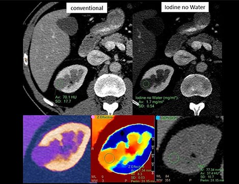
Case study: lesion characterization
The patient in this image was referred for characterization of a lesion in the kidney, suspected to be a kidney cyst when viewed on conventional CT. The cystic lesion is visible in the conventional contrast CT image (top left), but that gives little information about what it is. Switching to a visualization that enhances iodine and suppresses water (top right), the measurement indicates that there is iodine uptake and the lesion is metabolically active. Looking at the effective atomic number (bottom center), it can be seen that the Zeff value is 8.3, well above the value of water. The bottom right image is virtual non-contrast, filtering out all iodine signal and showing just the anatomy of the kidney. All spectral information in this case assisted in confirming this kidney malignancy, with just a single scan. In yet another view it is possible to check the heterogeneity of the lesion. Tumor heterogeneity is used as a guide to create more refined treatment strategies and response to therapy.
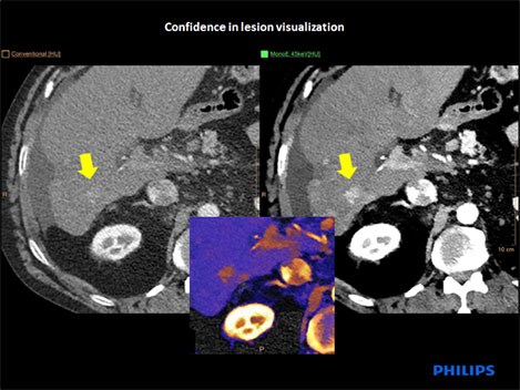
Case study: lesion visualization
This scan is a follow-up after ablation of hepatocellular carcinoma to see the effect of treatment. This patient also has a history of liver cirrhosis. On the conventional CT image (top left) there appears to be a poorly defined area of enhancement (yellow arrow). Changing to monoenergetic view at low energy (top right) boosts the iodine signal, showing that the area of the lesion is metabolically active. An overlap image of the iodine signal in color (center) clearly shows the de-novo hepatocellular carcinoma.
Now, the first exam is the right exam
With the Philips IQon Spectral CT, spectral results are available 100% of the time, in one scan. You can use them routinely for the most challenging cases and even apply insights from spectral data to incidental findings. No additional CT scans needed.
Acquisition of the spectral data is fully integrated with your current workflow, from scanner to PACS. An IQon Spectral CT scan always provides a traditional CT image – and if you need more information, you can just apply the spectral view retrospectively, from virtually anywhere. Spectral results are generally available in five minutes.
For more information
Visit IQon Spectral CT product page, click here.